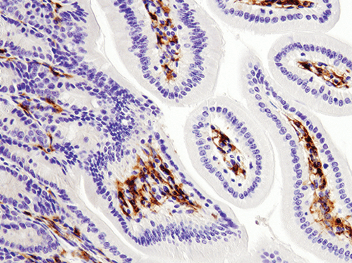
The preparation of samples for Immunohistochemistry (IHC) is a determining step in the process of these immunoassays; each tissue must be properly collected and prepared according to each study.
The preparation of these samples should be done according to the fixation method used, which in turn will depend on the detection technique chosen.
In this post we present you a brief guide with the keys to sample preparation for Immunohistochemistry.
The samples for immunohistochemistry can be prepared as sections of paraffin or frozen sections, do you know the difference?
The first of these would be in the fixing step . While fixation is carried out in the paraffin sections before the tissue is embedded in it, in the case of frozen sections the tissues are not fixed until after sectioning.
Another of the big differences is the conservation conditions of the tissues . While paraffin sections can be stored at room temperature for long periods of time, frozen sections do not usually remain stable for more than a year, and should always be kept at -80ºC.
Regarding the advantages and disadvantages of each of the techniques, we can highlight the following:
| ADVANTAGE | DISADVANTAGES | |
| PARAFFIN SECTIONS | Maintains the morphological characteristics of the tissues | Excessive fixation time could mask epitopes |
| FROZEN TISSUE SECTIONS | Maintains the enzymatic and antigenic functions of the tissues | Crystal formation could alter tissue structure |
Let’s see in more detail what the sample preparation procedure for immunohistochemistry would be in each case.
Paraffin-embedded tissue sections are often the choice when intending to preserve tissue samples for long periods of time.
Sample processing is faster in this case, but the stability of the samples is much less, so they cannot be stored for long periods of time.
On the other hand, in the case of frozen tissues, the sections are usually somewhat thicker, and may lead to poorer resolution under a microscope.